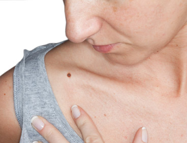
It is important to know what your moles look like. Most moles are harmless but skin cancer can develop in or near a mole. It can help to find and detect skin cancer earlier if you know what your moles look like.
Moles can be:
- One color – usually brown but can be tan, black, pink, blue, skin toned or colorless.
- Round or oval in shape
- Flat or slightly raised
- Look the same from month to month
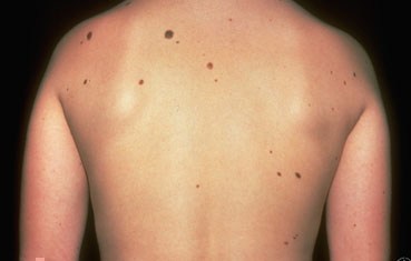
Moles can differ in size, shape or color. Moles can have hair. Some moles can change slowly over time, possibly even disappearing.
Nevus is the medical term for a mole. Nevi is the medical term for two or more moles.
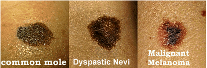
Types of Moles- Common or typical mole (nevus)
- Atypical mole (Dysplastic nevus) – This type can look like melanoma. It is not melanoma but you have a higher risk for melanoma if you have 4 or more dysplastic nevi.
- Congenital mole – This is when a person is born with a mole. About 1 out of every 100 people have congenital nevi. Having giant congenital nevi increases risks for melanoma.
- Spitz nevus – This often looks like melanoma. It can so closely resemble melanoma it can be difficult to determine whether or not it is a melanoma under the microscope.
- Acquired mole – When a mole appears after a person is born; greater than 50 increases risk for melanoma
Familial Atypical Multiple Mole-Melanoma Syndrome (FAMMM)
This is a genetic condition in which people have many moles (more than 50). Some are atypical. There is also a blood relative with melanoma. This increases your risk of developing melanoma
Other Benign Skin Lesions Seborrheic Keratoses (Seb Ks)
These common benign skin growths can occur anywhere on the skin. Some people have a few; others have many.
Most Seb Ks begin as a small rough bump on the skin. This tends to grow slowly, gradually thickening and often looks like a wart.
They are often brown but can range in color from light tan to black and can be various sizes.
They are distinguished by their waxy, pasted on the skin appearance. This makes it appear as though they can be easily removed with a fingernail.
It seems as though Seb Ks tend to run in families and sun exposure may play a role. Estrogen may also be a contributing factor. These lesions are more common with age.
Seb Ks are benign, so it is not absolutely necessary to remove these lesions. However, if a lesion grows quickly, turns black, itches or bleeds, it is typically removed. Lesions that are large or irritated may be removed as well.
Acrochordons (Skin Tags)These are fleshy growths that appear in the neck, groin, eyelids and under the arms. They can be skin colored to brown.
Genetics, friction and obesity all play a role in the development of skin tags. Skin tags may be a marker for insulin resistance.
Cherry Angiomas Dermatofibroma Solar Lentigenes Sebaceous Hyperplasia Keloid Cysts Milia Pilar Cysts Lipomas