Category Archives: Uncategorized

Psoriasis

psoriasis-plaque-psoriasis-vulgaris
Psoriasis and Psoriatic Arthritis
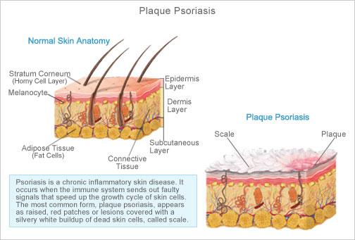
Psoriasis is an inflammatory skin condition that is typically chronic. It can vary from mild to very severe. It can be with or without joint pain.
Psoriasis is not contagious but can run in families.
Causes of Psoriasis


Exact cause is not known, but it is believed that the immune system plays a key role. The immune system activates T-cells (a type of white blood cell) and this causes the skin to grow too quickly. Skin typically replaces itself every 30 days, but in a person with psoriasis, the skin replaces itself about six times faster.
People typically have things that “trigger” psoriasis.
Common triggers:
  • Stress
  • Infection
  • Certain Medications
  • Cold, dry winter weather
  • Lack of sunlight
  • Injury – appears 10-14 days after

Types of Psoriasis
Psoriasis-types-pic

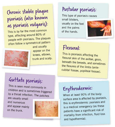
There are five major types of psoriasis:
 
  • Plaque Psoriasis

Most common type. Appears as patches of raised, red skin with silvery white scale. They are typically on the elbows, knees, lower back and scalp.
  • Guttate Psoriasis

These are small, red spots that usually affect children and young adults. Typically starts off as an infection.

  • Pustular Psoriasis

White pustules surrounded by red skin. Typically on the palms and soles.

  • Inverse Psoriasis


Smooth red lesions in skin folds.
  • Erythrodermic Psoriasis

Widespread redness with severe itching and pain. This can be life threatening.

Psoriatic Arthritis
Psoriatic arthritis develops in 10-30% of people with psoriasis. This is typically chronic joint pain. Medication can prevent joint problems if started early. Without treatment, permanent degeneration can occur
Treatment of Psoriasis
Psoriasis cannot be cured. Treatments can be very effective in controlling it, however.
Types of Treatment
  • Topicals

  • Corticosteroids
    Creams, ointments and lotions may clear the skin by reducing inflammation.
    Calcipotriene
    Form of vitamin D that controls growth of skin cells.
    Coal Tar

  • Light Therapy

  • Ultraviolet light slows rapid growth of skin cells

  • Systemic

  • Methotrexate
    This is an anti-cancer medication that can clear psoriasis. Regular blood tests are needed because of the effects of the medication on the liver.
    Retinoids
    This can cause dryness of the skin. It can be prescribed alone or in combination with UV light. Regular blood tests are needed.
    Cyclosporine
    This medication suppresses the immune system and is usually used to prevent organ rejection in patients who have received a transplant. Regular blood tests are needed.

  • Biologic

  • These medications are typically given by injection or infusion. They are unique in that they pinpoint the exact part of the immune system that is involved with psoriasis.
    There are many different biologic medications that may be used, given at different intervals.

Protect Your Skin

Protect-your-skin-from-the-sun

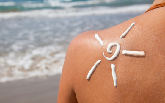
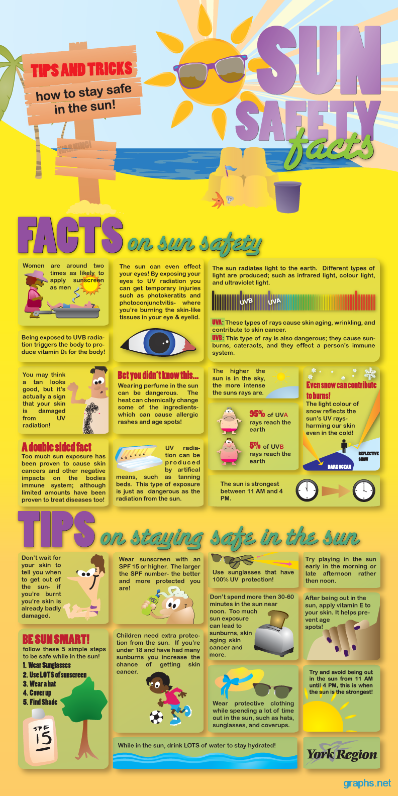
If you use common sense and take care to be sun smart, you can safely play and work outdoors without increasing skin cancer risk or premature aging.  It’s never too late to start protecting your skin.

  • Generously apply broad spectrum water resistant sunscreen with SPF 30 or more to exposed skin.  Re-apply every 2 hours, even on cloudy days, and after swimming or sweating.
  • Wear protective clothing, such as long sleeved shirts, pants, a wide brimmed hat and sunglasses, when possible.
  • Seek shade, when appropriate, remembering that the sun’s rays are the strongest between 10 am and 4 pm.  If your shadow is shorter than you are, seek shade.
  • Protect children from sun exposure by playing in the shade, wearing protective clothing and applying sunscreen.
  • Use extra caution near water, snow and sand, as they can reflect and intensify the damaging rays of the sun which can increase your chance of sunburn.
  • Get vitamin D safely through a healthy diet and include vitamin supplements as necessary. Don’t seek the sun.
  • Check your birthday suit on your birthday.  If you notice anything changing, growing or bleeding on your skin, have it checked.


melanoma-graphic  

summer-sun-safety-tips-and-facts

Moles

photo_2011_01_05_skin_cancer

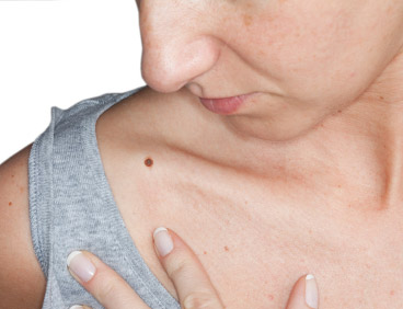
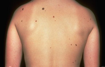
It is important to know what your moles look like.  Most moles are harmless but skin cancer can develop in or near a mole.  It can help to find and detect skin cancer earlier if you know what your moles look like.

Moles can be:

  • One color – usually brown but can be tan, black, pink, blue, skin toned or colorless.
  • Round or oval in shape
  • Flat or slightly raised
  • Look the same from month to month


Moles can differ in size, shape or color.  Moles can have hair.  Some moles can change slowly over time, possibly even disappearing.

Nevus is the medical term for a mole.  Nevi is the medical term for two or more moles.

Types of Moles

  • Common or typical mole (nevus)
  • Atypical mole (Dysplastic nevus) – This type can look like melanoma.  It is not melanoma but you have a higher risk for melanoma if you have 4 or more dysplastic nevi.
  • Congenital mole – This is when a person is born with a mole.  About 1 out of every 100 people have congenital nevi.  Having giant congenital nevi increases risks for melanoma.
  • Spitz nevus – This often looks like melanoma.  It can so closely resemble melanoma it can be difficult to determine whether or not it is a melanoma under the microscope.
  • Acquired mole – When a mole appears after a person is born; greater than 50 increases risk for melanoma


moles_nevi

Familial Atypical Multiple Mole-Melanoma Syndrome (FAMMM)

This is a genetic condition in which people have many moles (more than 50).  Some are atypical.  There is also a blood relative with melanoma.  This increases your risk of developing melanoma

atypical-moles

Melanoma

chart_image
Melanoma is the most serious form of skin cancer.  It is important to find and treat melanoma before it spreads.  With early diagnosis and treatment, melanoma has a high cure rate.

Causes of Melanoma

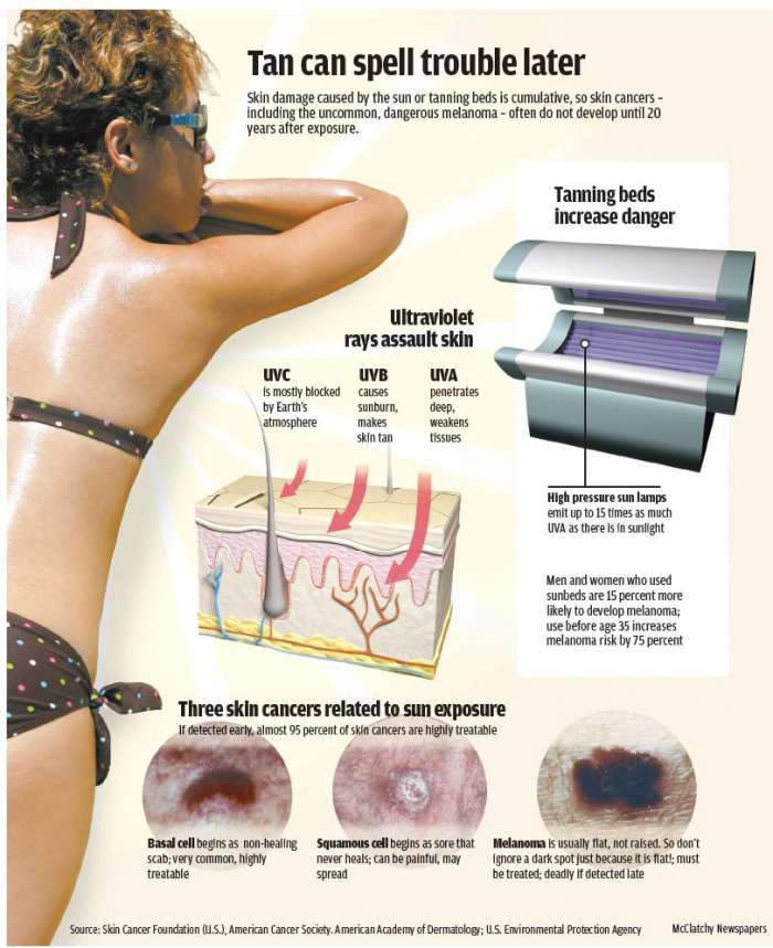
Exposure to UV rays plays a role in developing melanoma, especially in fair skinned people.  Many sunburns, especially blistering sunburns, raises the risk of getting melanoma.  Not all melanomas are caused by UV radiation. There are other risk factors that increase the likelihood of a person getting melanoma.

Risk Factors for Melanoma

  • Fair, sun sensitive skin that tans poorly or burns easily
  • Red or blond hair
  • Blue or green eyes
  • 50 or more small moles
  • Unusual looking moles that are often larger than normal and have uneven edges (may be called dysplastic nevi or atypical moles)
  • Past sunburns or indoor tanning
  • Past history of melanoma or other skin cancer
  • Blood relatives who have had melanoma
  • Weak immune system, due to disease, organ transplant or medicine
  • Age 50 or older


Being younger than 50 does not mean that you cannot get melanoma.  It is the most common cancer for young adults age 25-29 years old and the second most common cancer in adolescents and young adults.

Warning Signs of Melanoma

Nettedavi_Malign-Melanom-_496709

The most common warning sign of melanoma is change.  Melanoma may start in an existing mole.  A change in shape, color, diameter of a mole can be a sign of melanoma.  Other changes to watch out for are moles that begin to itch or bleed.

Not all melanomas start out in a mole.  Some melanomas begin suddenly on normal looking skin.  A sudden new growth could be melanoma.

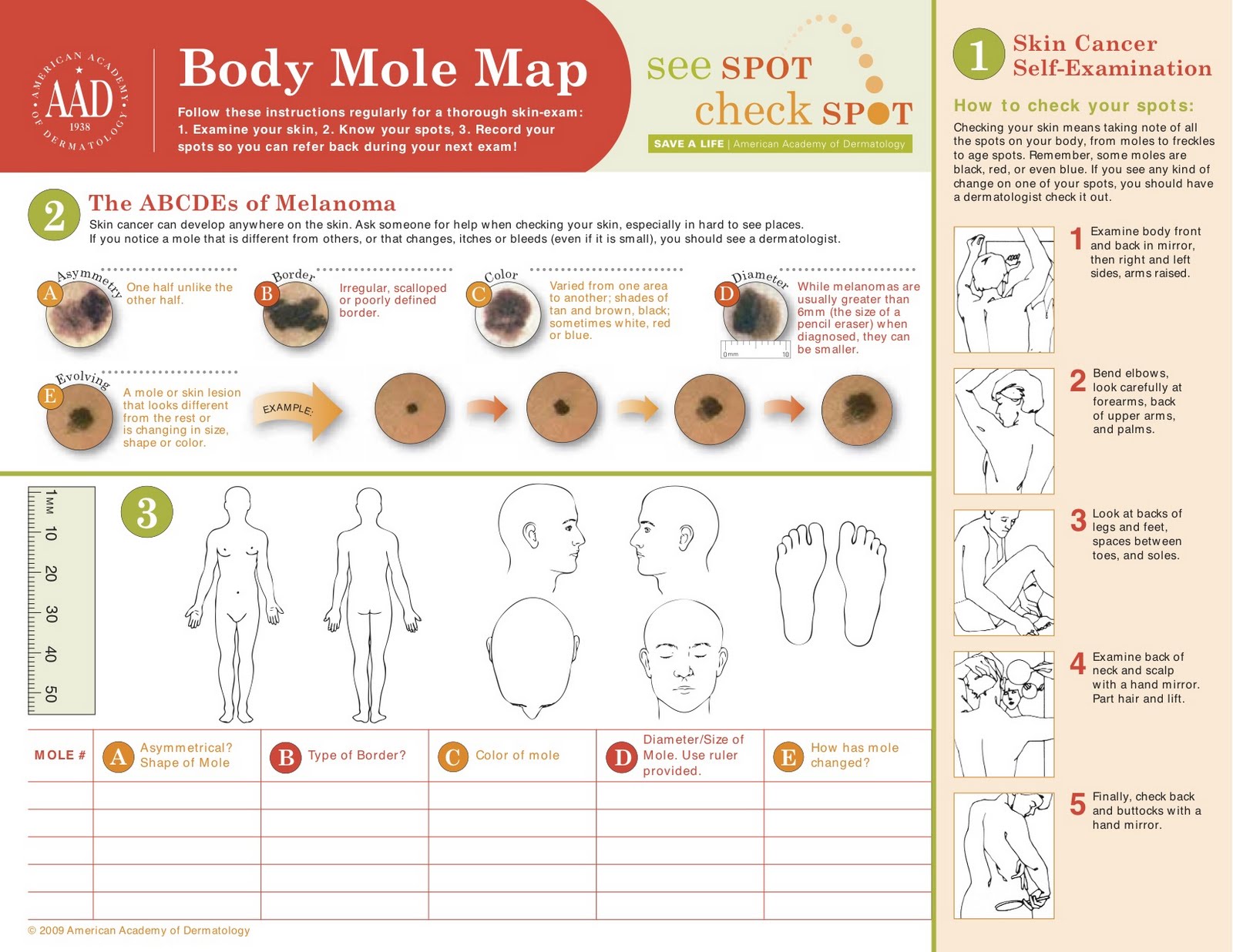
You should perform skin self exams, at least yearly, to look for warning signs.  It is helpful to look for the ABCDEs of melanoma detection.

A:  Asymmetry – one half does not look like the other half

B:  Border – border is irregular, scalloped or poorly defined

C:  Color – color is varied from one spot to another; there are shades of tan, brown and black; sometimes, red, white or blue

D: Diameter – melanomas are most often greater than 6 mm or the size of a pencil eraser (but they can be smaller)

E:  Evolving – A mole or growth that looks different from the rest or is changing in size, shape or color.

skincancer_abcde

The most likely place for melanoma to appear is on the upper back, torso, lower legs, face, scalp and neck, but can appear anywhere.  It can also begin under nails, inside the mouth or on the genitals, and even in your eye.

It is important to remember that melanomas can look different from each other, but the most significant observation is that they look different from your other moles.  You can often look for the “ugly duckling” – a mole that looks completely different than the other moles on your body.

If You Find a Mole That Concerns You

Make an appointment for a skin check.  If any of the moles are concerning, a biopsy will be performed.  During the biopsy, a very small needle is used to inject numbing medication into the skin surrounding the site for the procedure.  A sample of skin is then removed and sent to the pathologist for microscopic examination.  The pathologist will determine whether or not cancer cells are present and will typically tell what stage the cancer is.

Knowing the stage is important because different stages require different treatments.  To determine the stage, it is possible that you could have other testing performed such as ultrasound, x-ray, CT, MRI or PET scans.  Sometimes a surgical procedure known as a sentinel lymph node biopsy is necessary to determine the stage.  Near by lymph nodes are biopsied to determine whether or not the melanoma has spread.  Not all patients will need any or all of these procedures.

Stages of Melanoma

  •  Stage 0; in situ:  Melanoma is confined to the epidermis (top layer of skin)
  • Stage I-II:  Melanoma is confined to the skin, but has reached the second layer of skin (dermis) or beyond.
  • Stage III:   Melanoma has spread to nearby lymph nodes.
  • Stage IV:  Melanoma has spread to the internal organs, beyond the closest lymph nodes or to other lymph nodes or areas of the skin far from the first tumor.


compo_page1_big

Treatment of Melanoma

Treatment usually starts with excision (surgical removal) of the melanoma and some normal looking skin around it.  If taken early enough, this may be the only treatment necessary.  The sample is sent to the pathologist, who will determine whether or not cancer cells are in the surrounding skin.  If there are no cancer cells present, and the melanoma is in situ, cure rate is near 100%.

If the cancer has spread beyond the skin, more treatment will be needed.  This can be more surgery to remove the tumor and radiation or chemotherapy to kill cancer cells.  If melanoma is advanced, patients typically receive combination therapy.

Melanoma Can Return

Having melanoma increases your risk for developing new melanomas.  They can also recur or return.  Melanoma can spread.  Because of this, it is essential to keep all appointments and have regular skin checks along with self skin checks at home.

More Information

ABCDEs of Melanoma

Moles

The Sun and Your Skin

Performing Skin Self Checks
Mohs Micrographic Surgery

Mohs is a specialty surgical procedure that is not performed at this office.  If it is recommended that you have the Mohs procedure, you will need to go to a specialist for this type of procedure.

Physicians that perform Mohs are members of the American College of Mohs Surgery (ACMS) typically.  They are fellowship trained and have experience and expertise necessary to produce optimal outcomes in skin cancer treatment.

Areas that are difficult to treat with excision and radiation are typically recommended for Mohs because of the high cure rate and cosmetic outcomes.  This allows the smallest amount of tissue to be removed by surgeons trained in reconstruction surgery.

The Mohs Surgery Process

  • Step 1:  The roots of a skin cancer may extend beyond the visible portion of the tumor.  If these roots are not removed, the skin cancer will return.
  • Step 2:  The visible portion of the tumor is surgically removed.
  • Step 3:  A layer of skin is removed and divided into sections.  This is then color coded with dyes and a map of the surgical site is drawn.
  • Step 4:  The undersurface and edges of each section are microscopically examined for evidence of remaining cancer.
  • Step 5:  If cancer cells are found under the microscope, the surgeon marks the location on the map and returns to the patient to remove another layer of skin, but only precisely where cancer cells remain.
  • Step 6:  The removal process stops when there is no longer any evidence of cancer remaining in the surgical site.  Because Mohs surgery removes only tissue containing cancer, it ensures that the maximum amount of healthy tissue is kept intact.


For more information, please visit the ACMS patient website.

What to expect from a TCA peel

TCA peels are typically performed during the fall and winter months.  It is advised to avoid sun exposure following the peel as this can cause hyperpigmentation.  We typically start performing TCA peels in the clinic in November for this reason.

This type of peel typically requires a down time of 7-10 days. Skin will be red and swollen. Blisters may form and break open. This will not leave scars, however. Skin will scale and peel within 7-10 days. We recommend moisturizing the skin immediately after and several days after the peel.

The day of the peel:

Avoid wearing makeup the day of the peel. If makeup is worn, you will be asked to remove makeup and wash with a mild cleanser. TCA solution is then applied to dry skin. During the peel, your skin will feel tingly, prickly and then turn hot. There is usually a fan running to cool your skin and ease the burning sensation. “Frosting” sometimes occurs – your skin may turn white during the procedure. This may look scary, but this is the area where your skin will turn brown and peel away the most.

After the peel has completed, you will be asked to wash your face very gently and apply moisturizer. Do not use any abrasive products and do not scrub skin after a peel has been performed.

Days Following the Peel:

Skin will be red in the beginning and turn brown and begin to peel away. It may even blister and break open. This will not leave scars. Continue to use moisturizer and stay out of the sun. You can use makeup at this point as peeling has typically not yet begun.

Everyone responds differently to TCA peels. Some people will have more peeling and redness and some will have less. Peeling typically begins on day 3 and may even peel for 10 days. Do not pick at skin or peel away layers. This can cause damage. Continue to use moisturizer and stay out of the sun.

Antigens

The following is a list of antigens for which we test.

Dust

Feather Mix

  • Chicken Feathers
  • Goose Feathers
  • Duck Feathers

Cat Hair

Cattle Hair/Skin

Horse Hair/Skin

Cockroach

Dust Mites

Mold Mix 1

  • Alternaria alternata
  • Aspergillus fumigatus
  • Aspergillus glaucus
  • Aspergillus niger
  • Fusarium oxysporum
  • Helminthosporium sativum
  • Hormodendrum cladosporiodes
  • Mucor racemosus
  • Penicillum notatum
  • Phoma destructiva
  • Pullaria pullulans
  • Rhizopus nigricans

Mold Mix 2

  • Candida albicans
  • Epidermophyton floccosum
  • Trichophyton rubrum
  • Epicoccum nibrum
  • Botrytis cinerea
  • Fusarium vasinfectum
  • Trichophyton tonsurans
  • Chaetomium globosum

Environmentals

  • Cotton Linters
  • Kapok
  • Orris Root
  • Pyrethrum

Tree Mix 1

  • American Beech
  • American Elm
  • American Sycamore
  • Black Walnut
  • Black Willow
  • Eastern Cottonwood
  • Hard (Sugar) Maple
  • Red (River) Birch
  • Shagbark Hickory
  • White Ash

Tree Mix 2

  • Red Mulberry
  • Bald  Cypress
  • Hackberry
  • Black Locust
  • Sweetgum
  • Box Elder
  • Juniper

Oak Mix

  • Blackjack Oak
  • Bur Oak
  • Post Oak
  • Red Oak
  • White Oak

Pine Mix

  • Austrian Pine
  • Loblolly Pine
  • Scotch Pine
  • White Pine

Southern Grass Mix

  • Bermuda
  • Kentucky Blue
  • Orchard
  • Redtop
  • Timothy
  • Johnson
  • Fescue
  • Perennial Rye
  • Sweet Vernal

Weed Mix

  • Cocklebur
  • Lamb’s Quarters
  • Rough Pigweed
  • Sheep Sorrel
  • Yellow Dock
  • Marshelder
  • English Plantain
  • Goldenrod

Ragweed Mix

  • Giant
  • Short
  • Western

Grass Smuts

  • Bermuda
  • Johnson

Grain Smuts

  • Corn
  • Loose Barley
  • Loose Wheat
  • Oat

Mosquito