Monthly Archives: January 2014

Seborrheic Dermatitis

sebderm

Seborrheic Dermatitis (Seb Derm) is a common skin disease.  The rash that results is red and scaly.  It is typically found on the scalp, sides of the nose, eyebrows, ears, eyelids and middle of the chest.  It is easily treated, but is chronic, so it will recur if treatment stops.

Seb Derm is not dandruff.  Dandruff causes scaling on the scalp but not redness.  Seb Derm is characterized by scaling and redness.


Seb Derm is most often found in infants and older adults, but can affect anyone.  It is sometimes called “cradle cap” in infants.  In some infants, it can be only in the diaper area and be confused with diaper rash.

cradlecap4

If Seb Derm appears after infancy, it can be lifelong.  It can start off as dandruff and progress to full blown seborrheic dermatitis.

Seb Derm is more common in:
  • People who have a family member with seborrheic dermatitis
  • Men than women
  • People who have oily skin or hair
  • People with acne or psoriasis
  • People with Parkinson’s Disease or HIV/AIDS
  • People with alcoholism

Causes

Most doctors agree that the presence of a yeast like organism and excess of oil from the skin contribute to seborrheic dermatitis.  Immune system sensitivity is also a contributing factor.  Excess oil from skin is a food source for yeast to grow and the yeast causes inflammation, irritation and scaling.


Diagnosis

Most of the time, a diagnosis is made simply from looking at the rash on the body.  No special tests are required.  Seb derm that does not respond to treatment may need testing to rule out other diseases.

images

Treatment

There is no way to prevent or cure seb derm.  Treatment can control it, however.  It can also get better on its own.

Gentle shampooing with mild shampoo helps infants with cradle cap.  Low dose steroids or anti-fungals may also be applied.

Adults usually need a medicated shampoo and sometimes a steroid cream that is prescribed.

Protect Your Skin

Protect-your-skin-from-the-sun

If you use common sense and take care to be sun smart, you can safely play and work outdoors without increasing skin cancer risk or premature aging.  It’s never too late to start protecting your skin.


  • Generously apply broad spectrum water resistant sunscreen with SPF 30 or more to exposed skin.  Re-apply every 2 hours, even on cloudy days, and after swimming or sweating.
  • Wear protective clothing, such as long sleeved shirts, pants, a wide brimmed hat and sunglasses, when possible.
  • Seek shade, when appropriate, remembering that the sun’s rays are the strongest between 10 am and 4 pm.  If your shadow is shorter than you are, seek shade.
  • Protect children from sun exposure by playing in the shade, wearing protective clothing and applying sunscreen.
  • Use extra caution near water, snow and sand, as they can reflect and intensify the damaging rays of the sun which can increase your chance of sunburn.
  • Get vitamin D safely through a healthy diet and include vitamin supplements as necessary. Don’t seek the sun.
  • Check your birthday suit on your birthday.  If you notice anything changing, growing or bleeding on your skin, have it checked.


melanoma-graphic  

summer-sun-safety-tips-and-facts

Moles

photo_2011_01_05_skin_cancer

It is important to know what your moles look like.  Most moles are harmless but skin cancer can develop in or near a mole.  It can help to find and detect skin cancer earlier if you know what your moles look like.

Moles can be:

  • One color – usually brown but can be tan, black, pink, blue, skin toned or colorless.
  • Round or oval in shape
  • Flat or slightly raised
  • Look the same from month to month


Moles can differ in size, shape or color.  Moles can have hair.  Some moles can change slowly over time, possibly even disappearing.

Nevus is the medical term for a mole.  Nevi is the medical term for two or more moles.


Types of Moles

  • Common or typical mole (nevus)
  • Atypical mole (Dysplastic nevus) – This type can look like melanoma.  It is not melanoma but you have a higher risk for melanoma if you have 4 or more dysplastic nevi.
  • Congenital mole – This is when a person is born with a mole.  About 1 out of every 100 people have congenital nevi.  Having giant congenital nevi increases risks for melanoma.
  • Spitz nevus – This often looks like melanoma.  It can so closely resemble melanoma it can be difficult to determine whether or not it is a melanoma under the microscope.
  • Acquired mole – When a mole appears after a person is born; greater than 50 increases risk for melanoma


moles_nevi

Familial Atypical Multiple Mole-Melanoma Syndrome (FAMMM)

This is a genetic condition in which people have many moles (more than 50).  Some are atypical.  There is also a blood relative with melanoma.  This increases your risk of developing melanoma

atypical-moles

ABCDEs of Melanoma

abcdes-of-melanoma-large (1)  

Always get a pigmented lesion checked if there is any change at all.  There are typical things that you should look for in evaluating moles or lesions on the skin during self checks.  These are the ABCDEs.

A:  Asymmetry – One side is not like the other.  If you draw a line through the mole, the sides will not match.

B:  Border – The borders of an early melanoma may be uneven. The edges may be scalloped or notched.


C:  Color – Having a variety of colors is another warning signal. A number of different shades of brown, tan or black could appear. A melanoma may also become red, blue or some other color

D:  Diameter – Melanomas usually are larger in diameter than the size of the eraser on your pencil (1/4 inch or 6 mm), but they may sometimes be smaller when first detected.

E:  Evolution – Any change — in size, shape, color, elevation, or another trait, or any new symptom such as bleeding, itching or crusting — points to danger.

MELANOMA QUICK FACTS


Melanoma is one of the fastest growing cancers in the U.S. and worldwide. Consider:

  • One in 50 Americans has a lifetime risk of developing melanoma
  • People under 30 are developing melanoma at an alarming rate – the incidence soaring by 50% in young women since 1980
  • Melanoma is the most common form of cancer for young adults aged 25-29 & the second most common cancer in adolescents and young adults aged 15-29
  • In 2009, nearly 63,000 people were diagnosed with melanoma in the U.S., resulting in about 8,650 deaths
  • The American Cancer Society projects that nearly 77,000 will be diagnosed and 9,500 will diefrom melanoma this year
  • Every eight minutes, someone in the U.S. will be diagnosed with melanoma
  • Every hour of every day, someone will die from the disease

Actinic Keratosis

img-actinickeratosis

Actinic Keratoses (AKs) are very common. They are one of the most common reasons for office visits.  They are sometimes called solar keratoses because they are caused by years of sun exposure.

AKs are considered precancerous.  If they are left untreated there is a high likelihood of progression to squamous cell carcinomas

What Do AKs Look Like?

Most are dry, scaly and rough textured.  Some are skin colored and more difficult to see.  They are usually sandpaper like and are in groups that cover large areas of skin.  Some can appear as red bumps, thick red scaly patches or growths or crusted growths varying in color from red to brown to yellowish black.

Sometimes AKs grow rapidly upward from the skin and develop a growth that resembles a horn.  When this happens, the AK is called a cutaneous horn.  Horns vary in size and shape.  They are common on men’s ears.  They are typically considered cancerous as the base of the horn has a higher likelihood of cancer.

horn2

Treatment of AKs is important to prevent development into skin cancer.

Risk Factors For AKs

  • Blond or red hair color
  • Blue, green or hazel eyes
  • Skin that freckles or burns when in the sun
  • 40 years of age and older
  • Fair skin people
  • People who have had a lot of sun exposure
  • Using a tanning bed or sun lamp


Where Do AKs Form On The Body

AKs typically form in areas that receive the most sun.

  • Face, forehead and scalp, especially a bald scalp
  • Ears
  • Neck and upper chest
  • Back
  • Arms and hands
  • Lower legs, especially in women
  • Border of the lip – actinic chelitis


Actinic Chelitis:

Actinin-Cheilitis

Treatment Options

  • Cryosurgery:  This is the most common treatment.  This involves freezing the AK with liquid nitrogen, which will make the skin blister and flake off.
  • Chemical Peeling:  A chemical solution is applied to the sun to peel away the AK.
  • Chemotherapy for the skin:  A prescription called 4-fluorouracil can be written. This is a cancer fighting cream that you apply to the AK to destroy it.
  • Immunotherapy for the skin:  A prescription cream called imiquimod cream – works with the body’s immune system to help destroy AKs.
  • NSAIDs for the skin: Sodium diclofenac gel is a medication that destroys AKs.

Melanoma

chart_image
Melanoma is the most serious form of skin cancer.  It is important to find and treat melanoma before it spreads.  With early diagnosis and treatment, melanoma has a high cure rate.

Causes of Melanoma

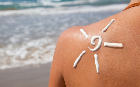
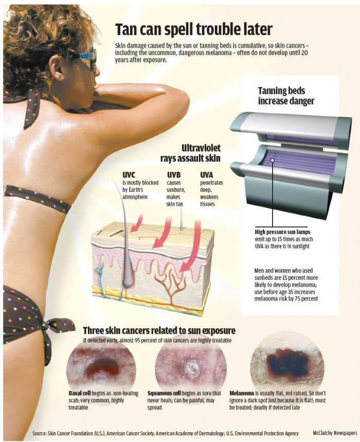
Exposure to UV rays plays a role in developing melanoma, especially in fair skinned people.  Many sunburns, especially blistering sunburns, raises the risk of getting melanoma.  Not all melanomas are caused by UV radiation. There are other risk factors that increase the likelihood of a person getting melanoma.

Risk Factors for Melanoma

  • Fair, sun sensitive skin that tans poorly or burns easily
  • Red or blond hair
  • Blue or green eyes
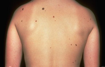
  • 50 or more small moles
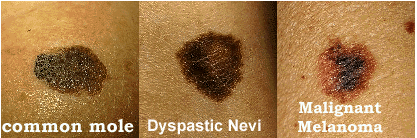
  • Unusual looking moles that are often larger than normal and have uneven edges (may be called dysplastic nevi or atypical moles)
  • Past sunburns or indoor tanning
  • Past history of melanoma or other skin cancer
  • Blood relatives who have had melanoma
  • Weak immune system, due to disease, organ transplant or medicine
  • Age 50 or older


Being younger than 50 does not mean that you cannot get melanoma.  It is the most common cancer for young adults age 25-29 years old and the second most common cancer in adolescents and young adults.

Warning Signs of Melanoma

Nettedavi_Malign-Melanom-_496709

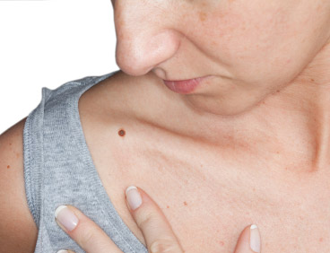
The most common warning sign of melanoma is change.  Melanoma may start in an existing mole.  A change in shape, color, diameter of a mole can be a sign of melanoma.  Other changes to watch out for are moles that begin to itch or bleed.

Not all melanomas start out in a mole.  Some melanomas begin suddenly on normal looking skin.  A sudden new growth could be melanoma.

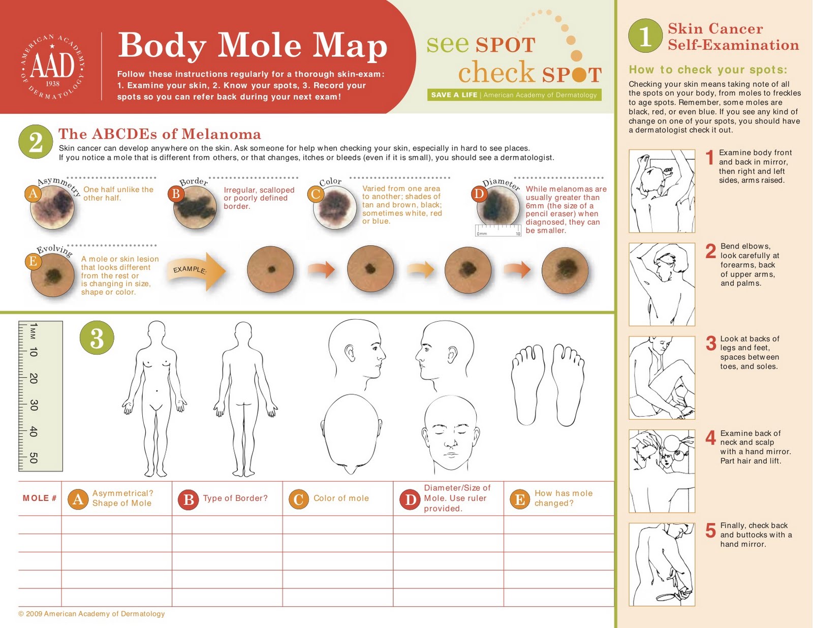
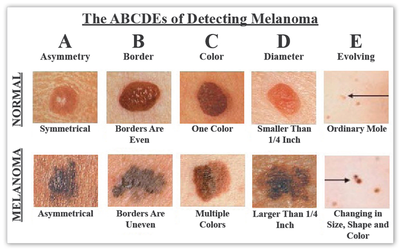
You should perform skin self exams, at least yearly, to look for warning signs.  It is helpful to look for the ABCDEs of melanoma detection.

A:  Asymmetry – one half does not look like the other half

B:  Border – border is irregular, scalloped or poorly defined

C:  Color – color is varied from one spot to another; there are shades of tan, brown and black; sometimes, red, white or blue

D: Diameter – melanomas are most often greater than 6 mm or the size of a pencil eraser (but they can be smaller)

E:  Evolving – A mole or growth that looks different from the rest or is changing in size, shape or color.

skincancer_abcde

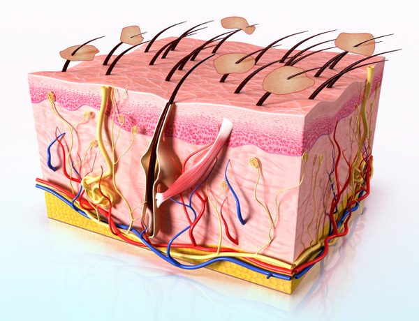
The most likely place for melanoma to appear is on the upper back, torso, lower legs, face, scalp and neck, but can appear anywhere.  It can also begin under nails, inside the mouth or on the genitals, and even in your eye.

It is important to remember that melanomas can look different from each other, but the most significant observation is that they look different from your other moles.  You can often look for the “ugly duckling” – a mole that looks completely different than the other moles on your body.

If You Find a Mole That Concerns You

Make an appointment for a skin check.  If any of the moles are concerning, a biopsy will be performed.  During the biopsy, a very small needle is used to inject numbing medication into the skin surrounding the site for the procedure.  A sample of skin is then removed and sent to the pathologist for microscopic examination.  The pathologist will determine whether or not cancer cells are present and will typically tell what stage the cancer is.

Knowing the stage is important because different stages require different treatments.  To determine the stage, it is possible that you could have other testing performed such as ultrasound, x-ray, CT, MRI or PET scans.  Sometimes a surgical procedure known as a sentinel lymph node biopsy is necessary to determine the stage.  Near by lymph nodes are biopsied to determine whether or not the melanoma has spread.  Not all patients will need any or all of these procedures.

Stages of Melanoma

  •  Stage 0; in situ:  Melanoma is confined to the epidermis (top layer of skin)
  • Stage I-II:  Melanoma is confined to the skin, but has reached the second layer of skin (dermis) or beyond.
  • Stage III:   Melanoma has spread to nearby lymph nodes.
  • Stage IV:  Melanoma has spread to the internal organs, beyond the closest lymph nodes or to other lymph nodes or areas of the skin far from the first tumor.


compo_page1_big

Treatment of Melanoma

Treatment usually starts with excision (surgical removal) of the melanoma and some normal looking skin around it.  If taken early enough, this may be the only treatment necessary.  The sample is sent to the pathologist, who will determine whether or not cancer cells are in the surrounding skin.  If there are no cancer cells present, and the melanoma is in situ, cure rate is near 100%.

If the cancer has spread beyond the skin, more treatment will be needed.  This can be more surgery to remove the tumor and radiation or chemotherapy to kill cancer cells.  If melanoma is advanced, patients typically receive combination therapy.

Melanoma Can Return

Having melanoma increases your risk for developing new melanomas.  They can also recur or return.  Melanoma can spread.  Because of this, it is essential to keep all appointments and have regular skin checks along with self skin checks at home.

More Information

ABCDEs of Melanoma

Moles

The Sun and Your Skin

Performing Skin Self Checks

Mohs Micrographic Surgery

Mohs is a specialty surgical procedure that is not performed at this office.  If it is recommended that you have the Mohs procedure, you will need to go to a specialist for this type of procedure.

Physicians that perform Mohs are members of the American College of Mohs Surgery (ACMS) typically.  They are fellowship trained and have experience and expertise necessary to produce optimal outcomes in skin cancer treatment.

Areas that are difficult to treat with excision and radiation are typically recommended for Mohs because of the high cure rate and cosmetic outcomes.  This allows the smallest amount of tissue to be removed by surgeons trained in reconstruction surgery.

The Mohs Surgery Process

  • Step 1:  The roots of a skin cancer may extend beyond the visible portion of the tumor.  If these roots are not removed, the skin cancer will return.
  • Step 2:  The visible portion of the tumor is surgically removed.
  • Step 3:  A layer of skin is removed and divided into sections.  This is then color coded with dyes and a map of the surgical site is drawn.
  • Step 4:  The undersurface and edges of each section are microscopically examined for evidence of remaining cancer.
  • Step 5:  If cancer cells are found under the microscope, the surgeon marks the location on the map and returns to the patient to remove another layer of skin, but only precisely where cancer cells remain.
  • Step 6:  The removal process stops when there is no longer any evidence of cancer remaining in the surgical site.  Because Mohs surgery removes only tissue containing cancer, it ensures that the maximum amount of healthy tissue is kept intact.


For more information, please visit the ACMS patient website.

Squamous Cell Carcinoma

EXAMPLE-SCC  

Squamous cell carcinoma (SCC) is the second most common type of skin cancer.  If this type of skin cancer is detected early, it can be cured with treatment.

What SCC looks like

SCC can appear in many different forms.  It  can be a bump that feels crusty and rough, a flat patch that is red and rough, a dome shaped bump that grows and bleeds or a sore that does not heal or that heals and returns.

Most of these types of skin cancer develop on sun exposed skin but they can develop in areas that are not exposed to the sun as well.

Some begin as a precancerous growth called an actinic keratosis (AK).  AKs are typically dry, scaly and rough.  They can be very small or even as large as a quarter.

Risk factors for SCC

Although SCC can develop on anyone with any skin type, there are certain risk factors that make it more likely for you to develop an SCC at some point in your life.  This is more common in caucasians.  UV radiation causes damage to the skin and makes it more likely that a skin cancer will develop.  As damage accumulates, risk increases.  Risk also increases with time because more exposure occurs over time.

The following is a list of risk factors for SCC:

  • Pale or light colored skin
  • Blue, green or gray eyes
  • Blond or red hair
  • Inability to tan
  • Significant exposure to UV light
  • Using tanning beds or sunlamps
  • Exposure to cancer causing chemicals
  • Smoked or used tobacco
  • Spending lots of time near heat, such as a fire
  • Previous diagnosis of AKs
  • Badly burned skin
  • Having an ulcer or sore on the mouth that has been there many years
  • Taking medicine that suppresses your immune system
  • Receiving an organ transplant
  • HPV infection
  • Receiving many x-rays
  • Many PUVA light treatments
  • Medical conditions – Xeroderma Pigmentosum, Epidermolysis Bullous or Albinism


Treatment Options for SCC

Early detection provides the best treatment options for SCC.  If it is not treated, it can spread to other areas of the body and make it more difficult to treat.

A biopsy must be performed to get an accurate diagnosis.  To have the biopsy performed, numbing medicine is injected into the skin surrounding the possible skin cancer.  A sample of tissue is obtained and sent to a pathologist to view under the microscope.

The diagnosis given by the pathologist will determine treatment options.  The options available depends on how deep the skin cancer is, how large it is, and whether or not it has spread to other areas.

  • Excision – Surgical procedure performed in office.  This is performed in the same fashion as the biopsy, with local injections of numbing medicine.  The area of skin containing skin cancer, along with normal looking skin is taken off and sent to the pathologist to look at under the microscope.  The pathologist will determine if any cancer cells are in the normal looking skin.  If there are no cancer cells, then we say that the margins are clear and no further treatment is needed.  If there are cancer cells present, more treatment will be needed.
  • Mohs Micrographic Surgery – Surgical procedure performed in the office under local anesthesia.  This means that injections are given into the skin and you will be awake for the procedure.  The surgeon will remove the area of skin that is affected by cancer and a small amount of normal looking skin.  He or she will then look at the sample under the microscope immediately to determine whether or not margins are clear.  If they are not clear, more skin is taken and the process is repeated until all margins are clear.
  • Radiation – When a person cannot undergo surgery to treat a skin cancer, they may be sent for radiation therapy.  A series of radiation treatments are used to destroy the cancer cells.
  • Medicine applied to the skin – If caught early enough, a prescription medication can be given to be applied to the skin at home to destroy cancer cells.
  • Cryotherapy – For superficial SCC, those just on the surface of the skin, liquid nitrogen can be used to destroy cancer cells.  A very cold air is applied to the skin, usually by spraying from a canister to “freeze” the cancer cells to destroy them.

Basal Cell Carcinoma

EXAMPLE-BCC-S  

BCC is the most common form of skin cancer.  Millions are diagnosed each year and nearly all are cured with treatment.

What BCC Looks Like

  • Dome shaped growth with visible blood vessels
  • Shiny, pinkish patch
  • Sore that heals, then returns and repeatedly heals and returns
  • Brown or black growth
  • White or yellow waxy growth that looks like a scar


Most BCCs develop on sun exposed skin such as the scalp, neck and hands.  It is especially common on the face – on the nose, cheeks and forehead but can develop anywhere on the body.

Risk Factors For BCC

Anyone can get BCC, but there are risk factors that make it more likely that you will be diagnosed with BCC at some point in your life.  Lighter skinned people tend to develop BCC more frequently.  Damage to the skin occurs through UV exposure, either from the sun or from tanning beds.  Over time, this damage accumulates and increases the risk of developing skin cancer.  The risk does increase with age because the older you are, the longer you have been exposed to UV radiation.

Risk Factors:

  • Pale, light colored or freckled skin
  • Blond or red hair
  • Blue, green or gray eyes
  • Family history of skin cancer
  • A weakened immune system or are taking medications that suppress the immune system
  • Received radiation therapy
  • Used tanning beds or other indoor tanning devices


BCC is serious because it can grow deep enough to affect bone or other tissue.  It rarely spreads to other areas of the body, however.  A deep or invasive BCC can be difficult to treat.

If caught and treated early, BCC is highly curable.  Treatment cures most BCCs.  To diagnose a BCC a biopsy will be performed.  This is the only way to diagnose any type of skin cancer.  The biopsy will be performed under local anesthesia, which means that the area of skin to be biopsied will be numbed with injections of lidocaine.  The tissue sample will then be sent to a pathologist to be examined under a microscope.  If the diagnosis is BCC, there are many factors that will determine the appropriate treatment.

Treatment Options for BCC

  • Excision – Surgical procedure to remove the BCC along with normal looking skin to ensure that all of the cancer cells have been removed.  This is sent to the pathologist for confirmation of clear margins, which means that there are no remaining cancer cells in that tissue that remains.  If the normal looking skin contains cancer cells, more treatment is necessary.
  • Mohs micrographic surgery – Mohs is usually performed at a medical office under local anesthesia while you remain awake.  The skin cancer, along with some normal looking skin is removed.  The sample is then examined under the microscope to determine whether or not cancer cells remain, and where those cells are.  This will direct the surgeon as to where to remove more skin sample, if necessary.  This process continues until there are no cancer cells remaining.
  • Radiation – When surgery cannot be performed, radiation therapy may be recommended.  A series of radiation treatments are performed to destroy the cancer cells.
  • Medication applied to the skin – When BCC is caught very early and is superficial, medication may be applied to the skin at home to destroy cancer cells.